▲Researchers from NYCU, Academia Sinica, and France’s Institut Curie identify the TTBK2/HUWE1 regulatory pathway. This breakthrough, published in Cell Death & Differentiation, offers a new roadmap for treating pediatric brain cancer with significantly fewer side effects © NYCU Elite
In its early stages, “medulloblastoma” typically presents no obvious symptoms. As the tumor gradually grows, it obstructs cerebrospinal fluid flow, causing young children to experience vomiting and fainting due to elevated intracranial pressure. Often diagnosed only after emergency hospital admission, medulloblastoma is a notorious killer in pediatric wards. This malignant brain tumor that is prevalent in children, often requires emergency surgery to remove the tumor and save the pediatric patient’s life. However, subsequent treatment involving multiple courses of radiation therapy and chemotherapy to eliminate residual cancer cells frequently leads to severe sequelae such as cognitive impairment, motor function damage, and secondary cancers.
Finally, a decade-long multinational research collaboration between National Yang Ming Chiao Tung University, Academia Sinica, and Institut Curie in France has unraveled the regulatory mechanisms behind the abnormal proliferation of medulloblastoma cancer cells. This breakthrough offers a novel pathway for developing targeted therapies with reduced side effects, and it is expected to decrease the frequency and dosage of radiation therapy and chemotherapy for pediatric patients while significantly improving their prognosis and quality of life. This major research breakthrough has been published in the prestigious international journal Cell Death & Differentiation.
Root Out the Cause and Halt Tumor Proliferation at Its Root
During cerebellar development, neural stem cells first generate a small cluster of “granule neuron progenitors”. These progenitors continuously divide and proliferate after receiving growth signals through their “primary cilia” structures. When granule neuron progenitors retract their primary cilia, they cease division and prepare to differentiate into mature neurons. The research team has discovered for the first time that the proteins TTBK2 and HUWE1 play critically important roles in the process of primary cilia retraction.
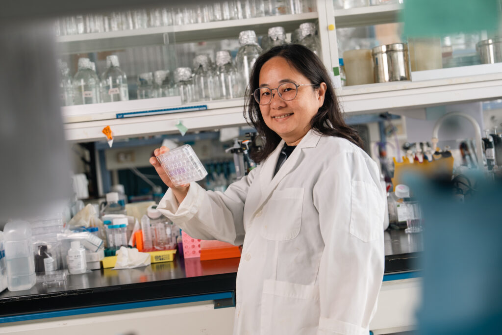
Professor Won-Jing Wang at NYCU Institute of Biochemistry and Molecular Biology described: “TTBK2 is like the ‘building permit’ for primary cilia growth. Once TTBK2 reaches the centrosome of the progenitor, it begins constructing cilia, receiving growth signals, and then proceeds with division and proliferation. When the progenitor prepares to differentiate into a mature neuron, HUWE1 is responsible for degrading TTBK2. Once the ‘building permit’ is revoked, the primary cilia retract. The progenitor stops dividing and starts to differentiate.”
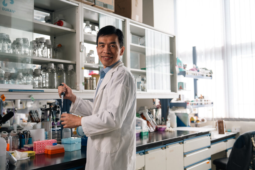
“However, in SHH-type medulloblastoma, this delicate dynamic equilibrium is disrupted!” Professor Jin-Wu Tsai at NYCU Institute of Brain Science indicates: “TTBK2 in cancer cells cannot be normally degraded. Primary cilia fail to retract and continuously receive growth signals, ultimately causing progenitors to become abnormally activated and uncontrollably proliferated, forming malignant tumors akin to ‘illegal structures’.”
After confirming TTBK2 as a key therapeutic target, the team is actively developing strategies to inhibit TTBK2. One approach involves developing small-molecule RNA drugs that interfere with TTBK2 mRNA, directly blocking TTBK2 production. Another approach focuses on preventing TTBK2 from reaching the centrosome. A major advantage of such targeted therapies lies in their “specificity”: mature cerebellar neurons typically lack primary cilia, so inhibiting TTBK2 affects only aberrantly proliferating tumor cells, significantly reducing side effects on normal brain tissue.
A Decade of Dedication to Mastery to Decode the Black Box of Cancer
This groundbreaking achievement did not happen overnight, but rather took root in a Taiwan-France collaborative research project that began in 2013. At that time, Olivier Ayrault, a cancer specialist from Institut Curie in France, visited Taiwan seeking partners for joint research on the molecular mechanisms underlying the formation of cerebellar medulloblastoma. Jin-Wu Tsai recalls: “NYCU Brain Science Laboratory has long focused on studying brain structure and developmental processes. Medulloblastoma is intrinsically linked to cerebellar development, making the collaboration a perfect fit.” Subsequently, Professor Won-Jing Wang, whose expertise lies in cell biology, biochemistry, and research on centrosomes and primary cilia, joined the team, completing the final piece of the puzzle in understanding the microscopic mechanisms.
This research truly brought together masters from various fields, each showcasing their unique expertise. Institut Curie is already a hallowed institution in tumor research, while the NYCU team possesses the “cerebellar electroporation” technique—a capability found in only a handful of laboratories worldwide. This technology enables precise gene transfer into the mouse cerebellum, allowing observation of how specific genes influence tumor growth. Moreover, NYCU’s cutting-edge neuroimaging technology allows researchers to clearly visualize minute cellular changes. Academician Bon-chu Chung of Academia Sinica further validated the findings using zebrafish models, confirming that this regulatory mechanism exhibits high evolutionary conservation—from fish to mammals.
“This is the power of interdisciplinary collaboration.” Won-Jing Wang emphasized: “It’s like observing a black box—a single discipline can only see one side. By combining neuroscience’s macro perspective, cell biology’s micro mechanisms, and clinical oncology’s pathological analysis, we can piece together the full picture of pathogenesis.” Jin-Wu Tsai added with deep resonance that as communication technology advances, it has made cross-border, interdisciplinary collaboration both more vital and accessible. Jin-Wu Tsai said: “This collaboration model sparks cutting-edge research breakthroughs, thereby infinitely expanding the frontiers of research.
In addition to the long-standing collaboration with Institut Curie in France, Professor Jin-Wu Tsai is currently engaged in multinational research with a Belgian team. His recent findings on brain development and genetic mutations are set to be published in Nature Communications. These frequent and in-depth international connections fully demonstrate that NYCU’s research capabilities in the field of brain science have earned high recognition from the global academic community.
See the Bigger Picture through Small Details and Explore New Frontiers in Brain Science
In the past, scientists believed primary cilia were vestigial structures akin to the appendix, mere remnants of evolutionary processes. Today, they have been proven to be crucial hubs for progenitors to receive growth signals. The multinational research team led by Professors Won-Jing Wang and Jin-Wu Tsai has not only revealed the dual role of primary cilia in cerebellar development and tumorigenesis but also demonstrated the profound strength of NYCU in basic science and translational medicine. Their work offers new hope for life for pediatric patients lying on sickbeds.
However, the research team’s exploration does not stop here. Won-Jing Wang pointed out that recent academic findings suggest primary cilia appear to be highly correlated with drug resistance and malignancy in various cancers: “We have observed cilia with unusual shapes and varying lengths in certain highly malignant tumors or drug-resistant cancer cells. This may be the next field worthy of attention and investment in research.”
Additionally, the team’s research scope has expanded from “cell proliferation” to encompass “neurodegeneration” and “developmental abnormalities”. Jin-Wu Tsai said that mutations in the TTBK2 gene are known to be associated with cerebellar ataxia type 11. Meanwhile, mutations in centrosome proteins frequently cause brain developmental abnormalities, such as lissencephaly—a condition characterized by the absence of folds on the brain’s surface. Therefore, this collaborative effort will further investigate how these microscopic organelles influence the macroscopic structure and function of the brain, unraveling more unsolved mysteries in the field of brain science.
Interview | Fu-Kuo Chu
Translation | Yi-Chen Emily Li
Editing | Hsiu-Cheng Faina Chang / StoryLab
Photographer | Hao-Yun Peng and Yen-Yu Shih / ZDunemployed studio
©NYCU ELITE